
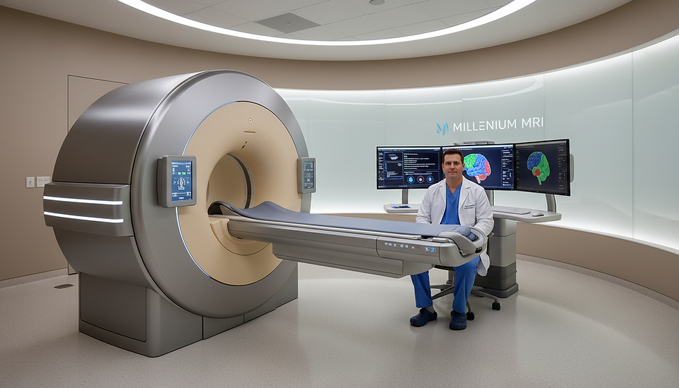
NeuroQuant MRI at Millenium MRI
- trieumri
- Feb 24
- 1 min read
Millennium MRI offers an advanced option that adds objective measurement to routine brain imaging. NeuroQuant software works with an MRI to quantify brain structure volumes, helping clinicians spot changes tied to dementia, injury, or other concerns.
The service is patient-centered and clear. It explains what the software adds, who benefits, and how the process moves from scan to report. Millennium MRI locations in Ocala, FL; Largo, FL; Tamarac, FL; Jonesboro, AR; Marion, AR; and North Little Rock, AR may offer this option. Availability varies by site.
What to expect: a comfortable scan, automated volume measurements, and a structured report that supports clinical decisions. The goal is clearer, measurable information when cognitive decline or injury is a concern.
Safety basics: an MRI uses no X-rays or ionizing radiation. For scheduling, the local team can confirm the correct exam based on the provider order and set an appointment.
Key Takeaways
NeuroQuant adds automated, objective brain volume measurements to imaging.
It helps track changes linked to dementia, injury, and cognitive decline.
Millennium MRI offers this service at multiple Florida and Arkansas locations.
Scans are safe; no ionizing radiation is used.
Scheduling teams verify exam availability and confirm your appointment.
NeuroQuant MRI: advanced brain imaging for objective brain volume measurements
An extra scan sequence provides automated volume data that complements routine brain imaging. This does not replace the standard exam; it adds a quantitative layer to help clinicians interpret results.
What the software adds to a standard brain mri
The software performs automated volumetric analysis on the acquired images. It measures specific regions and converts those images into numeric measurements clinicians can track over time.
What “brain structures” and “brain volume” mean in your results
Brain structures are named regions, such as the hippocampus and other areas often tracked in memory concerns. Brain volume is the measurable amount of tissue in those regions, reported as a number and a percentile compared to peers.
Measurements reduce ambiguity by turning visuals into objective data.
Quantitative results can reveal subtle changes not obvious on visual review.
The exam remains a standard mri at its core, with added analysis for the referring provider.
"Objective volume data can help clarify changes when symptoms like memory loss appear."
How NeuroQuant software analyzes brain structures and detects changes over time
Software tools extract precise volumes from scan images to give objective information about brain structures. This automated analysis produces consistent measurements for key regions, turning visual images into numeric data that clinicians can track.
Automated volumetric measurements
The program segments images and calculates volumes for each brain structure. Results are repeatable, so follow-up scans yield comparable numbers for monitoring progression.
Normative database comparison
Each volume is compared to an FDA-approved database adjusted for age, sex, and cranial volume. This context shows whether a brain structure’s size is typical or below expected for that age and sex.
Tracking atrophy and progression
Reports flag patterns of age-related atrophy and quantify changes between exams. Clinicians evaluate whether volume loss is stable, gradual, or rapid—information that affects management and follow-up.
Metric | Typical rate | Clinical note |
Hippocampus shrinkage | ~1% per year (healthy) | Small shrinkage can be age-related |
Hippocampus shrinkage | ~5% per year (Alzheimer disease) | Faster loss linked to higher progression risk |
Database comparison | Age/sex/cranial volume adjusted | Provides percentile and context |
"Smaller hippocampus size is associated with a higher likelihood of progression to Alzheimer disease in the next few years."
Clinical caution: this analysis supports assessment and monitoring, but diagnosis requires the provider’s full clinical judgment and other information.
When providers order a NeuroQuant brain MRI scan
Clinicians order a quantitative brain scan when they need reproducible numbers to guide diagnosis and follow-up. This add-on provides objective volumetric data to support evaluation, monitoring, or treatment planning for neurological concerns.

Alzheimer’s disease evaluation and monitoring
Alzheimer disease evaluation often uses volumetric measures to spot patterns of regional atrophy. Measured change in areas like the hippocampus helps clinicians track progression over time rather than relying on symptoms alone.
Dementia workups and memory loss concerns
For dementia and memory loss, imaging helps narrow possible causes. Quantitative results are one piece of information used with clinical exams, labs, and history to identify likely conditions.
Mild Cognitive Impairment as a potential transition stage
When mild cognitive impairment is suspected, repeated scans can show whether structural decline is accelerating. Tracking numbers over time may help providers decide on follow-up or interventions.
Traumatic brain injury and brain injury-related cognitive decline
After traumatic brain injury, quantitative imaging can document structural impact and support recovery assessment. Objective volumes may help monitor worsening or improvement in conjunction with clinical care.
Clinical note: the referring provider determines if this scan is appropriate based on symptoms, history, and prior results. Imaging informs care but does not by itself define diagnosis.
Benefits of quantitative brain imaging data for diagnosis and treatment planning
Measured brain volumes provide a factual basis that supports diagnosis and treatment planning. Objective numbers reduce uncertainty and make findings easier to discuss with patients and families.
Supporting earlier, more confident clinical decisions
Objective measurements add clarity to visual reads. When imaging shows subtle change, numeric data can prompt earlier evaluation or referral.
Monitoring whether decline is accelerating
Serial volume measurements reveal trends. Tracking numbers across exams shows if changes are steady or accelerating, which may alter follow-up plans.
Helping assess treatment effectiveness over time
Quantitative reports let clinicians compare results before and after an intervention. Stable or improving volumes may support continuing a plan; worsening results can trigger adjustments.
Use | What it shows | Clinical value |
Early detection | Small regional change | Earlier diagnostic workup |
Progress monitoring | Trend across exams | Assess acceleration or stability |
Treatment assessment | Pre/post volume comparison | Guide therapy decisions |
Contextual interpretation | Age-adjusted atrophy metrics | Differentiates expected aging from concerning patterns |
"Quantitative data complements clinical judgment and helps tailor care for varied conditions."
NeuroQuant MRI vs standard brain MRI imaging
Routine imaging captures detailed brain pictures; additional software turns those pictures into trackable data.
What a standard brain mri shows: A conventional exam gives a radiologist high-resolution images to spot lesions, bleeding, tumors, and structural change. It relies on visual assessment and descriptive reporting.
What quantitative volumetric analysis adds: The same images are processed by software to produce numeric measurements of specific brain structures. These volumes are compared to norms and prior scans so clinicians can see subtle change over time.
How they work together
Standard scans deliver anatomy and signal findings that guide immediate diagnosis.
Quantitative analysis provides repeatable measurements to support monitoring and research.
The software does not replace the radiologist; it complements human interpretation with structured information.
Measured outputs are especially useful when symptoms are subtle or when a baseline is needed for future comparison.
Aspect | Standard brain mri | Quantitative analysis | Clinical value |
Primary output | Visual report and images | Numeric volumes and percentiles | Combines look and numbers for fuller context |
Use case | Acute findings, lesions, structural detail | Tracking atrophy, subtle change over time | Better trend detection and objective follow-up |
Who interprets | Radiologist | Radiologist with software report | Human judgement informed by measurements |
Patient takeaway | Shows what the brain looks like | Shows how specific parts measure and change | Both approaches are clinically valuable |
"Standard MRI shows what the brain looks like; NeuroQuant helps quantify how specific parts measure and change."
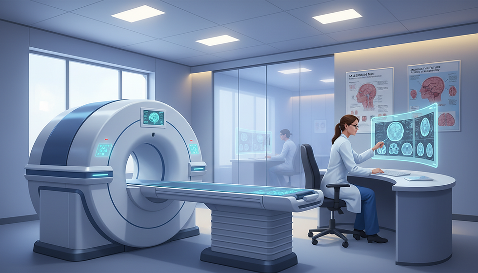
What to expect during your MRI scan at Millennium MRI
A focused, supportive workflow helps patients move smoothly from arrival to completion of the brain scan.
Check‑in and preparation: At arrival, staff confirm the physician order and review safety questions. Patients change if needed and remove metal or jewelry. Technicians answer questions and explain timing so the appointment feels predictable.
NeuroQuant as an added scan sequence
The neuroquant software sequence is added to a routine brain mri exam. Patients should expect the same basic experience; the extra sequence simply provides measured volume data and adds a few minutes to the scan.
MRI with and/or without contrast: how providers decide
Contrast is used only when a provider believes it will help answer the clinical question. The team reviews history, allergies, and kidney health before any contrast is given.
MRI safety basics: no X‑rays or ionizing radiation
Safety note: the scan uses magnetic fields, not X‑rays. Screening for implants or metal in the body is essential to protect patient safety.
During the scan: patients lie on a padded table that moves into the scanner while images are acquired. Staff remain in contact and can provide ear protection or comfort aids for anxiety.
Step | What to expect | Typical time |
Check‑in | Registration, safety review, change | 10–20 min |
Scan | Padded table, images acquired, extra sequence added | 20–40 min |
Contrast (if used) | IV placement, brief observation | 5–10 min |
Aftercare | Technician review; resume normal activity | 5–10 min |
"Share any concerns about comfort or past exams so the team can support a smoother appointment."
Preparing for your NeuroQuant MRI appointment
Knowing what to bring and what to avoid makes the appointment smoother and helps staff capture clear, usable images. Patients who arrive prepared reduce delays and get better data for their provider.
What to bring
Physician order and ID.
Any prior images or reports that relate to the reason for the scan.
Insurance card and a list of current medications.
What to wear and leave at home
Wear comfortable clothing without metal fasteners. Remove jewelry, watches, and hair clips. Leave valuables at home when possible; lockers may be available at the facility.
Medication and day‑of reminders
Continue prescribed medication unless the ordering provider gives other instructions. Some sites advise avoiding food or drink for two hours before the scan; confirm with the scheduling team.
Special considerations
Disclose implants, pacemakers, artificial valves, or any metal in the body. Mention pregnancy or if nursing so staff can plan for safety. Share any known conditions that could affect the appointment.
"Bring prior reports and tell the team about implants or pregnancy to keep the visit safe and efficient."
Understanding your NeuroQuant report and results
Patients receive a single, organized report that shows what the images reveal and how key regions measure compared to norms.
How neuroradiologists interpret image findings and volumetric data
A neuroradiologist reviews both the visual scan and the neuroquant software analysis. They integrate measurements and image findings to form one clinical interpretation.
That synthesis helps the referring provider see whether observed changes match symptoms, history, or expected aging.
How results are delivered to your referring provider
The final report includes standard mri findings plus the volumetric analysis and structured measurements. Results are sent to the ordering physician.
The provider reviews the report with the patient, explains the information, and recommends next steps when needed.
How reports support ongoing monitoring for dementia and other conditions
Serial reports document changes and patterns of brain atrophy over time. Repeated measurements help track progression of dementia or other neurodegenerative disease.
Bring prior studies for comparison; trends often offer the most meaningful clinical insight.
"The report is designed to support medical decisions and guide follow-up; it does not replace a full clinical evaluation."
What the report contains | Why it matters | Patient action |
Visual findings | Shows lesions, signal changes, structural detail | Discuss with referring provider |
Volumetric data and measurements | Provides numeric comparison to norms | Save prior results for trend analysis |
Integrated radiologist interpretation | Combines images and data into one conclusion | Follow recommended next steps |
Schedule NeuroQuant MRI near you: locations, hours, and contact information
Call the local scheduling team to set an appointment for the scan using your physician order. The staff will confirm exam availability and help pick a convenient time.

Ocala, FL
2023 E Silver Springs Blvd Unit 301, Ocala, FL 34470
P: (352) 900-5501 | F: (352) 900-5502
Hours: Monday–Friday 9am to 5pm; Saturday & Sunday Closed
Jonesboro, AR
2929 South Caraway Road, Ste. 6, Jonesboro, AR 72401
Contact numbers are not listed for this site. Patients should call Millennium MRI central scheduling or a nearby location for assistance with booking the scan.
Marion, AR
2860 I 55, Suite 8, Marion, AR 72364
P: (870) 275-7749 | F: (870) 275-6073
Largo, FL
2900 East Bay Drive, Largo, FL 33771
P: (727) 683-6501 | F: (727) 683-6503
Tamarac, FL
7201 N. Pine Island Road, Tamarac, FL 33321
P: (954) 720-0903 | F: (954) 720-4583
North Little Rock, AR
800 W. 4th St., North Little Rock, AR 72114
P: (501) 500 0051 | F: (501) 500 0052
Before you call: have your provider order ready. Ask about available appointment times, any prep specific to your health, and whether contrast may be used.
"Bring your order and prior reports so the scheduling team can book the correct exam and advise what to bring."
The team can confirm arrival instructions and answer common questions to make the visit smooth. Call today to schedule your mri appointment and secure the scan that fits your needs.
Conclusion
Pairing high-quality brain imaging with software-driven analysis turns pictures into repeatable numbers clinicians can trust.
The NeuroQuant option adds automated volumetric quantification and FDA-database comparison to a standard MRI scan. Those objective results support clearer discussion about dementia, memory loss, and other cognitive concerns.
A neuroradiologist integrates the software output with visual review and sends the organized report to the referring provider for next-step planning and diagnosis guidance.
Patients should schedule the correct mri at Millennium MRI and bring their physician order and prior images. Early, accurate information helps caregivers and providers make more informed choices and plan follow-up calmly.
FAQ
What is NeuroQuant MRI at Millennium MRI?
NeuroQuant MRI at Millennium MRI is an advanced brain imaging service that adds automated volumetric analysis to a routine brain scan. It provides objective measurements of brain structure sizes to help clinicians evaluate atrophy, injury, and disease progression.
What does the software add to a standard brain scan?
The software provides automated segmentation and volume measurements of key brain structures. These objective data supplement visual reads, giving clinicians precise numbers to track changes over time and compare results to population norms.
What do “brain structures” and “brain volume” mean in the results?
“Brain structures” refer to anatomical regions such as the hippocampus, cortex, ventricles, and subcortical nuclei. “Brain volume” indicates the size of those regions, usually reported in cubic centimeters and as percentiles versus an age- and sex-matched reference group.
How does the software analyze brain structures and detect changes over time?
The system uses automated algorithms to segment images and calculate volumes for each structure. When prior studies are available, the tool compares timepoints to quantify atrophy rates and flag accelerating decline.
Are measurements compared to a normative database?
Yes. Volumes are compared to an FDA-cleared normative database that accounts for age, sex, and cranial size. This comparison helps determine whether a structure is smaller than expected for someone of similar demographics.
How is age-related brain atrophy reported and tracked?
Reports present absolute volumes and percentile rankings, and they calculate changes between studies. Clinicians use these metrics to distinguish normal aging from pathological atrophy and to monitor progression.
Why is the hippocampus important for Alzheimer’s disease risk assessment?
The hippocampus plays a key role in memory. Shrinkage of this structure often appears early in Alzheimer’s disease and can suggest increased risk or early neurodegeneration when seen alongside clinical symptoms.
When would a provider order this type of brain scan?
Providers may order it for memory concerns, dementia workups, suspected Alzheimer’s disease, mild cognitive impairment, or to evaluate cognitive changes after traumatic brain injury.
How does quantitative brain imaging support diagnosis and treatment planning?
Quantitative data enable earlier, more confident clinical decisions by providing objective evidence of structural change. They also help clinicians monitor whether decline is accelerating and assess treatment effectiveness over time.
What does a standard brain scan show compared to volumetric analysis?
A standard scan shows structural detail and lesions on visual inspection. Volumetric analysis adds precise, repeatable measurements of region sizes and automated comparisons to norms, improving the objectivity of follow-up assessments.
What should I expect during my scan at Millennium MRI?
The volumetric sequence is added to a routine brain study and typically adds only a few minutes to scan time. Technologists will explain the process, and the exam is performed like a standard head scan.
Is contrast used, and how do providers decide?
Contrast is used only when clinically indicated, such as to evaluate inflammation, tumor, or blood–brain barrier breakdown. The referring provider and radiologist determine if contrast is necessary based on the clinical question.
Is the scan safe? Does it use radiation?
The scan uses strong magnetic fields and radio waves; it does not use ionizing radiation such as X-rays. Safety screening is performed to address implants, pacemakers, or other contraindications.
What should I bring to my appointment?
Bring the referring physician’s order and any prior brain images or reports. These help radiologists compare current and past studies for accurate tracking.
What should I wear and leave at home?
Wear comfortable clothing without metal fasteners. Leave jewelry, watches, credit cards, and other metal objects at home or in your vehicle to avoid interference with the scan.
Any medication guidance or day-of-scan reminders?
Continue prescribed medications unless instructed otherwise. Arrive on time, follow any fasting instructions if given, and inform staff about claustrophobia so accommodations can be arranged.
Are there special considerations for implants, pacemakers, pregnancy, or nursing?
Inform staff about implants, pacemakers, or if you are pregnant or breastfeeding. Some devices may be contraindicated; alternatives or additional safety steps may be required.
How do neuroradiologists interpret the volumetric report?
Neuroradiologists integrate volumetric measurements with the visual appearance of the brain and clinical history. They highlight significant deviations from norms and any lesion or structural abnormalities relevant to the referral.
How are results delivered to my referring provider?
Final reports and images are sent electronically to the referring provider. The physician will review findings with the patient and recommend next steps for diagnosis, monitoring, or treatment.
How can these reports support ongoing monitoring for neurodegenerative conditions?
Serial volumetric reports provide objective trends that help clinicians detect early progression, evaluate therapy response, and adjust care plans for conditions such as Alzheimer’s disease and other dementias.
Where can I schedule a scan at Millennium MRI?
Appointments are available at multiple locations, including Ocala, FL; Jonesboro, AR; Marion, AR; Largo, FL; Tamarac, FL; and North Little Rock, AR. Contact the nearest center for hours and booking details.


Comments