
MRI and multiple sclerosis: how MRI tracks disease progression
- trieumri
- Sep 24, 2025
- 1 min read

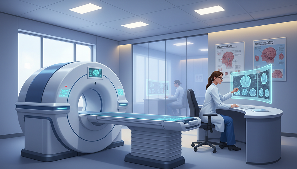
Magnetic imaging offers a clear view of the brain and spinal cord to confirm diagnosis, evaluate alternatives, and set a baseline when not contraindicated. A quality scan uses at least a 1.5 Tesla magnet; 3 Tesla is preferred to show fine detail and early changes.
This guide explains how imaging reveals inflammation, scarring, and subtle changes that may not show on exam. Readers learn which images clinicians take, what they reveal in white matter and other key regions, and how scans guide treatment choices over time.
Expect a practical approach to the first visit, timing of scans, and how results are explained to each patient. For hours and access: Monday-Friday, 9am to 5pm; Saturday & Sunday, Closed. For appointments and referrals, see Section 4 for Florida and Arkansas locations and contact numbers.
Key Takeaways
High-quality imaging sets a baseline that guides future care decisions.
Scans show disease activity in brain and spinal pathways not obvious on exam.
Standard protocols improve comparisons across time for early detection.
Contrast may be used selectively to answer specific diagnostic questions.
Results are shared in plain language to support safety and daily function.
Core principles of MRI in multiple sclerosis: what the images reveal and why they matter
High-resolution magnetic scans reveal where the nervous system has been injured and how those changes track over time. These images let clinicians see white matter changes and small lesions in the brain and spinal cord that relate to symptoms. Clear pictures also document the initial burden of disease so care teams can measure change.
How magnetic resonance detects lesions
Magnetic resonance techniques provide contrast between tissue types to highlight lesion locations in white matter pathways. Scans of the cervical and thoracic cord pick up cord lesions that often affect mobility, sensation, or balance.
Baselines, magnet strength, and follow-up
Baseline studies should include brain plus cervical and thoracic spinal cord, with and without contrast when safe. At least 1.5 tesla is recommended; a 3 tesla scanner improves detail in the cord and reduces artifact. Follow-up scans on the same magnet and protocol ensure apples-to-apples comparisons.
Images document lesion burden and guide early diagnosis and treatment planning.
Cord and posterior fossa findings raise conversion risk in radiologically isolated cases.
Consistent protocols improve sensitivity for small changes over time.
MRI and multiple sclerosis in diagnosis and monitoring: sequences, timing, and safety
Careful selection of sequences, timing, and safety checks ensures scans answer practical clinical questions.
Standard brain protocols prioritize 3D sagittal T2 FLAIR, 3D T2, 2D axial diffusion-weighted imaging, and 3D T1 MPRAGE. Axial T1 post-contrast is added when active inflammation is suspected. If 3D is unavailable, optimized 2D FLAIR and fast spin echo T2 are acceptable for diagnosis and follow-up.
Spine studies use sagittal T2, STIR, and T1 with axial T1/T2 to localize cord lesions that affect walking or hand function. Re-baseline at about 6 months after starting disease-modifying therapy, then schedule scans every 6–12 months based on activity and therapy type.
Contrast and safety: Macrocyclic gadolinium is preferred when needed; check renal function per guidelines. Non-contrast resonance imaging is preferred in pregnancy; breastfeeding may continue after contrast.
PML and therapy surveillance: High-risk patients require more frequent monitoring with FLAIR and DWI; post-contrast images are used selectively.
Consistency: Use the same magnet, matched protocols, and slice orientations to detect new lesions and small change over time.
Appointments run Monday–Friday, 9am–5pm, with Florida and Arkansas sites available for routine scans, contrast-enhanced studies, and safety screenings; see Section 4 for contact details.
From imaging to care decisions: interpreting new lesions and guiding treatment over time
Scan-detected changes offer objective evidence that guides timely adjustments in therapy and monitoring.
Clinicians combine lesion location, enhancing activity, and overall burden to estimate long-term risk. They weigh brain and spinal cord findings with exam results to form a clear prognosis.
When mri shows new lesions or growth despite treatment, the team reviews adherence and tolerance. They then consider escalation or a different therapy to protect function and quality of life.
Translating MRI data into prognosis: lesion burden, enhancing activity, and disability risk
Contrast-enhancing activity often signals recent inflammation, but non-enhancing T2 changes can still mean breakthrough disease. Cord lesions carry higher disability risk, so targeted scans are used even when symptoms are mild.
When MRI changes prompt therapy adjustments or escalation
Review adherence, side effects, and prior response before changing therapy.
Use comparable scans over time to confirm that a true event has occurred.
Increase monitoring frequency or add contrast when recent inflammation is suspected.
Patient access and scheduling: contact, hours, and locations in Florida and Arkansas
All sites are open Monday-Friday, 9am–5pm. Weekend appointments are closed.
Location | Address | Phone | |
Ocala, FL | 2023 E Silver Springs Blvd Unit 301, Ocala, FL 34470 | (352) 900-5501 | |
Largo, FL | 2900 East Bay Drive, Largo, FL 33771 | (727) 683-6501 | |
Tamarac, FL | 7201 N. Pine Island Road, Tamarac, FL 33321 | (954) 720-0903 | |
Jonesboro / Marion / North Little Rock, AR | Jonesboro: 2929 S Caraway Rd Ste 6; Marion: 2860 I 55 Suite 8; N Little Rock: 800 W. 4th St. | Jonesboro/Marion: (870) 275-7749 | North Little Rock: (501) 500 0051 |
Conclusion
Consistent scans on the same magnet make small changes visible and actionable, helping teams turn images into clear next steps.
Baseline studies that include brain plus cervical and thoracic spinal cord at 1.5 tesla or higher set a reliable reference. Standardized sequences and matched magnets improve detection of new or enlarging lesions and guide timely therapy changes.
When contrast is needed, macrocyclic gadolinium is preferred; non-contrast exams remain safe in pregnancy, and breastfeeding can continue after contrast. For prompt scheduling during weekday hours, contact the nearest Florida or Arkansas site listed above. Teams coordinate imaging, results, and follow-up so patients receive clear, timely care.
FAQ
What does magnetic resonance imaging show about brain and spinal cord disease?
Magnetic resonance imaging reveals lesions in white matter and the spinal cord that reflect active inflammation or older injury. Images highlight lesion size, location, and whether new or enhancing changes are present. Radiologists use these findings to estimate lesion burden and potential risk of future disability.
How does scanning detect white matter versus spinal cord lesions?
Different pulse sequences emphasize tissue contrasts. FLAIR highlights periventricular and cortical white matter lesions, T2 shows overall lesion load, and spinal sequences target the cervical and thoracic cord. Combining sequences gives a clearer picture of disease distribution across brain and cord.
Why is establishing a baseline scan important?
An initial scan sets a reference for future comparisons. Knowing preexisting lesions helps clinicians spot new events, measure progression, and make informed treatment choices. Guidelines recommend imaging both brain and cervical/thoracic cord during the first assessment.
Does magnet strength affect image quality and why is 1.5 Tesla considered the minimum?
Higher field strength improves signal, contrast, and lesion detection. A 1.5 Tesla scanner provides adequate clinical information, but 3 Tesla offers better sensitivity for smaller or subtle lesions, aiding more reliable longitudinal comparisons.
What is radiologically isolated syndrome and how do scans help?
Radiologically isolated syndrome refers to incidental findings of white matter lesions in people without symptoms. Serial imaging helps determine whether lesions remain stable or new activity emerges, which informs risk assessment and consideration of early interventions.
Which sequences are standard for brain and spine imaging?
Standard protocols include FLAIR and T2 for lesion detection, diffusion-weighted imaging for acute changes, and 3D volumetric scans for brain volume and atrophy measures. Spine protocols use sagittal and axial T2 and STIR or proton density to capture cord lesions accurately.
How often should follow-up scans be performed after diagnosis?
Many clinicians obtain a follow-up study at about six months to re-establish baseline, then repeat imaging every six to twelve months depending on disease activity, treatment changes, and clinical stability. Frequency increases with new symptoms or concern for progression.
When should the spinal cord be scanned even if there are no symptoms?
Asymptomatic cord lesions can carry prognostic significance. Scanning the cervical and thoracic cord at baseline and during follow-up helps detect silent disease that may influence prognosis and treatment strategy.
When is gadolinium contrast helpful during a scan?
Gadolinium highlights active, enhancing lesions and helps differentiate new inflammatory activity from older damage. Contrast is most useful during suspected relapse or when atypical features raise diagnostic uncertainty. Clinicians weigh benefit against safety considerations, especially with repeated exposure.
Are there special considerations for pregnancy, breastfeeding, or implanted devices?
Scanning without contrast is generally safe in pregnancy when clinically necessary; contrast use is reserved for pressing indications. Breastfeeding patients can usually continue nursing after contrast exposure. Implanted devices require device-specific safety checks and vendor guidance before scanning.
How does imaging support therapy surveillance and safety monitoring?
Regular imaging detects new T2 lesions or enhancing activity that may signal treatment failure and prompt therapy adjustment. For patients on certain immunomodulators, scans also monitor for rare complications such as progressive multifocal leukoencephalopathy, helping balance benefit and risk.
Why is consistency across scans important for long-term monitoring?
Using the same magnet strength, comparable protocols, and consistent slice orientation ensures valid comparisons over time. Apples-to-apples scans reduce false impressions of change and improve confidence in decisions about escalation or de-escalation of care.
How do clinicians translate imaging findings into prognosis?
Radiologists and neurologists combine lesion burden, presence of enhancing lesions, and patterns of atrophy to estimate future disability risk. Imaging is one piece of the puzzle alongside clinical exams, functional testing, and patient-reported symptoms.
When do imaging changes prompt therapy adjustments?
New or increasing lesion burden, repeated enhancing activity, or radiographic progression despite treatment often prompt consideration of therapy escalation. Decisions also factor in relapse frequency, side effects, and patient goals.
How can patients schedule scans and access care in Florida and Arkansas?
Patients should contact local imaging centers or neurology clinics for appointments. Offices typically list contact numbers, hours, and locations online. Referrals from treating neurologists or primary care providers often speed scheduling and ensure proper protocols are used.

Comments