
Multiple sclerosis MRI at Millenium MRI
- trieumri
- Feb 24
- 1 min read
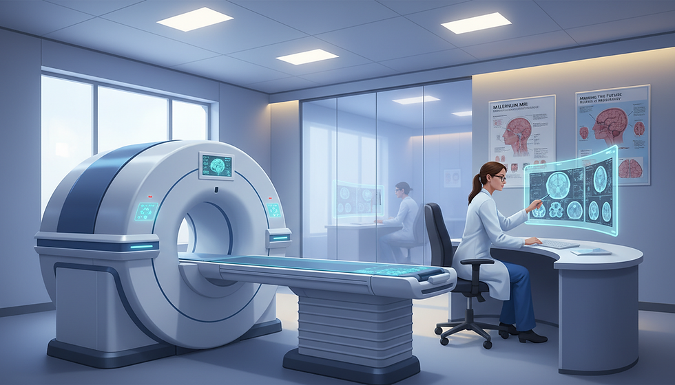
Millennium MRI offers focused care for people seeking answers about demyelinating disease. This guide explains what a multiple sclerosis MRI is meant to show and why tailored scan protocols matter for clear results.
The scan is a main tool to detect, diagnose, and monitor disease, used alongside clinical history and a neurologic exam. MRI sensitivity for diagnosis exceeds 90%, but other white matter conditions can mimic findings, so imaging must be read in clinical context.

This how-to overview covers what radiologists look for, why lesion pattern and location matter more than a simple spot count, and how results support accurate follow-up. It also previews when imaging is recommended and how the McDonald criteria use dissemination in space and time.
Appointments can be scheduled at Millennium MRI locations in Florida and Arkansas. As a convenience note, sites generally operate Monday–Friday, 9am to 5pm, and are closed Saturday and Sunday; full location details and phone numbers appear later in the article.
Key Takeaways
Dedicated scan protocols improve diagnostic clarity and follow-up.
Imaging is one piece of diagnosis; symptoms and exam matter too.
"Lesions" are areas of abnormal signal; pattern and site are key.
Millennium MRI emphasizes patient comfort and clear next steps.
Sites in Florida and Arkansas offer weekday hours for easy booking.
When a Multiple sclerosis MRI is recommended for symptoms and suspected multiple sclerosis
When new neurologic symptoms appear, timely brain and spine imaging helps guide next steps.
Common symptoms that prompt referral include vision changes such as pain with eye movement or blurred vision, numbness or tingling in limbs, balance and coordination problems, and subtle cognitive shifts that affect daily tasks.
Clinicians often order scans after a first neurologic event to evaluate for lesions that suggest inflammatory demyelination in the central nervous system. Prompt imaging can clarify whether findings raise concern for a longer diagnostic workup.
Understanding CIS and RIS
Clinically isolated syndrome is a first, single episode lasting at least 24 hours without fever or infection. An MRI at that time helps estimate the risk of later developing multiple sclerosis and informs follow-up plans.
Radiologically isolated syndrome describes an incidental scan that looks highly like MS in someone without clear symptoms. This usually triggers closer monitoring and targeted testing rather than an immediate diagnosis.
Why MRI is preferred over CT
CT is fast and useful in emergencies to rule out bleeding. For suspected demyelination, however, magnetic resonance imaging offers far better visualization of white matter and the spinal cord.
Use case | CT | MRI (dedicated protocol) |
Emergency bleed suspected | Fast, widely available | Not ideal first choice |
Detecting white matter lesions | Poor sensitivity | High sensitivity, better detail |
Spinal cord evaluation | Limited detail | Preferred for cord lesions |
Follow-up and treatment planning | Not recommended | Standard for monitoring |
Practical takeaway: patients with concerning symptoms should contact their clinician for an order, bring a clear symptom history, and arrange to have prior imaging sent to the center. Ask whether both brain and spine scans are being considered to support accurate diagnosis and planning.
How MRI helps with diagnosis multiple sclerosis using the McDonald criteria
Clinicians use targeted imaging plus clinical evidence to confirm whether typical lesion patterns meet formal diagnostic rules.

Objective evidence matters
The McDonald criteria require objective proof of lesions that are separated by both space and time, and exclusion of more likely causes. Doctors combine imaging with history, a neurologic exam, and other tests like CSF analysis or evoked potentials when needed.
Dissemination in space
Dissemination in space means lesions appear in two of four typical areas: periventricular, juxtacortical/intracortical, infratentorial, and spinal cord. Meeting this part of the criteria depends on location and appearance, not simply any white matter change.
Dissemination in time
Dissemination in time shows lesions formed at different moments. This can be proved by new T2 lesions on follow-up or by finding both enhancing and non‑enhancing lesions on the same scan. Gadolinium contrast highlights active inflammation; enhancement usually lasts about one month and helps estimate lesion age.
Patient tip: bring prior reports and images so radiologists can compare studies and confirm whether new lesions have appeared over time.
What radiologists look for on a Multiple sclerosis MRI
Careful pattern recognition — not counting spots — guides radiologists when interpreting scans for suspected demyelination. They review the full distribution across white matter, the gray–white junction, and the spinal cord to see whether findings are distinctly typical.
Periventricular lesions and Dawson fingers
Periventricular lesions that touch the ventricles and ovoid Dawson fingers aligned perpendicular to the ventricle surface point toward an inflammatory process. These track along small vessel pathways and are best seen when they are in firm contact with the ventricular margin.
In older adults or those with cardiovascular risk, radiologists often look for at least three periventricular lesions before calling the pattern supportive of sclerosis.
Juxtacortical, corpus callosum, and temporal lobe
Juxtacortical lesions must touch cortex and often involve U‑fibers; U‑fiber involvement helps separate MS‑like changes from common subcortical white matter changes.
Corpus callosum and temporal lobe lesions increase confidence in the diagnosis, though temporal lobe findings are interpreted in clinical context because other conditions can give similar images.
Brainstem and spinal cord features
Brainstem lesions from MS tend to be peripheral and asymmetric, unlike central, symmetrical small vessel changes. Spinal cord lesions are usually short (under two vertebral segments), peripheral, and often cervical — a pattern that adds specificity when seen with supportive brain findings.
Contrast timing
Gadolinium highlights active inflammation. Enhancement commonly lasts about one month. Imaging the T1 post‑contrast after roughly 15 minutes often improves detection depending on the facility's protocol.
How to avoid misdiagnosis: MS vs common white matter diseases and red flags
Not every bright spot on a scan signals the same disease; pattern and context guide correct labeling. Avoiding misdiagnosis protects patients from needless long-term therapy, anxiety, and missed alternative diagnoses.

MS-like patterns versus small vessel changes
Distinctive patterns — callosal, juxtacortical/U-fiber, temporal lobe, brainstem and short spinal cord lesions — increase confidence in an inflammatory diagnosis. In contrast, small vessel ischemic change often spares U‑fibers, looks symmetric, and appears with lacunes and vascular risk factors.
When to think beyond an inflammatory diagnosis
NMOSD: long cord lesions spanning >3 vertebral segments and central gray involvement.
ADEM: diffuse, often symmetric lesions with deep gray matter in a monophasic illness.
CADASIL: anterior temporal pole or external capsule findings plus migraine/family history.
Sarcoid: linear leptomeningeal or perivascular enhancement.
PML: subcortical U‑fiber/cortical involvement, DWI rim activity in immunosuppressed patients.
Checklist-based reading
Confirm lesions meet strict MS-like definitions (Dawson finger contact with ventricle; juxtacortical touches cortex).
Match imaging with exam and risk factors.
If only nonspecific white matter lesions are present, avoid labeling as demyelination without further workup.
Practical advice: ask for neuroradiology review and neurologist correlation when reports are vague. When MRI sensitivity is high but pattern is unclear, a second opinion or follow-up imaging improves diagnostic accuracy.
How to prepare for your MRI scan and what to expect on exam day
Arriving ready with prior studies and a concise symptom timeline helps the radiology team focus the protocol for best results.
Before you arrive: complete paperwork, bring or upload past imaging, and prepare a short history of symptoms and any medication list. Note prior contrast reactions and share them with staff.
With and without contrast
Some exams use images both with and without contrast to show dissemination time. Gadolinium is often given early; post-contrast T1 images may be acquired about 15 minutes later to better show active versus older lesions.
Comfort and image quality tips
Technologists position a head or spine coil depending on the referral. Staying still is key to clear imaging; short breaks and cushions can help.
The spinal cord protocol often uses PD or STIR sequences because spinal FLAIR shows few cord lesions. If both brain and spinal imaging are ordered, expect a longer appointment.
Follow-up expectations
Clinicians may plan repeat scans to detect new changes over time. Intervals depend on disease activity, therapy, and new symptoms. Ask about claustrophobia support or scheduling adjustments when you book.
Millennium MRI locations in Florida and Arkansas to schedule your appointment
Millennium MRI maintains several convenient sites across Florida and Arkansas to help patients schedule timely neuroimaging. Timely access supports accurate diagnosis and follow-up when neurologic concerns arise.
Please schedule using your clinician’s order, and confirm whether the request is for brain, spine, or both. Ask if contrast is included and request that prior images and reports be sent before the visit for direct comparison.
Bring ID and insurance, and arrive early for check-in. This helps the team begin protocol-specific scanning without delay and improves the chance of a useful comparison study.
All Location Hours
All Location Hours: Monday-Friday: 9am to 5pm | Saturday & Sunday: Closed
Site directory
Location | Address | Phone | Fax |
Ocala, FL | 2023 E Silver Springs Blvd Unit 301, Ocala, FL 34470 | P: (352) 900-5501 | F: (352) 900-5502 |
Jonesboro, AR | 2929 South Caraway Road, Ste. 6, Jonesboro, AR 72401 | — | — |
Marion, AR | 2860 I 55, Suite 8, Marion, AR 72364 | P: (870) 275-7749 | F: (870) 275-6073 |
Largo, FL | 2900 East Bay Drive, Largo, FL 33771 | P: (727) 683-6501 | F: (727) 683-6503 |
Tamarac, FL | 7201 N. Pine Island Road, Tamarac, FL 33321 | P: (954) 720-0903 | F: (954) 720-4583 |
North Little Rock, AR | 800 W. 4th St., North Little Rock, AR 72114 | P: (501) 500 0051 | F: (501) 500 0052 |
For a smoother visit, patients should confirm the exact mri protocol with the ordering clinician. Forward prior studies when possible to support accurate comparison and faster diagnosis. Staff can advise on contrast needs and expected appointment length.
Conclusion
A focused imaging exam gives clinicians measurable evidence to pair with symptoms and neurologic findings. For a reliable diagnosis, radiologic patterns, lesion distribution, and timing must fit the clinical picture to reduce false positives and to rule out other causes of disease.
Careful interpretation helps separate inflammatory disease from common mimics. Follow-up scans and complementary testing often clarify uncertain findings and support treatment planning.
For next steps, patients can use the site directory above to contact Millennium MRI in Florida or Arkansas, confirm Monday–Friday hours, and arrange sending prior studies for comparison.
FAQ
When is a scan recommended for symptoms that suggest demyelinating disease?
Imaging is advised when patients report new vision changes, unexplained numbness, balance or coordination problems, or sudden cognitive changes. These symptoms may reflect inflammatory lesions in the central nervous system and warrant timely evaluation to guide treatment.
What do clinically isolated syndrome and radiologically isolated syndrome mean for imaging?
Clinically isolated syndrome describes a first clinical episode consistent with inflammatory demyelination, and a focused scan helps assess lesion distribution. Radiologically isolated syndrome means lesions typical of inflammatory disease are seen incidentally on imaging without symptoms; such findings usually prompt neurologic follow-up and periodic imaging.
Why is magnetic resonance preferred over CT for detecting demyelination in the brain and spinal cord?
Magnetic resonance provides superior soft-tissue contrast and higher sensitivity for detecting small white matter and cord lesions. It visualizes lesion characteristics, locations, and enhancement patterns that CT cannot, making it the standard choice for evaluation.
How does imaging support diagnosis using the McDonald criteria?
Scans provide objective evidence of lesion number, location, and activity. When combined with clinical history, neurologic exam, and lab tests such as cerebrospinal fluid analysis, imaging helps demonstrate dissemination in space and time required by diagnostic criteria.
What is meant by dissemination in space on imaging?
Dissemination in space refers to lesions appearing in typical regions of the brain and spinal cord, such as periventricular areas, juxtacortical zones, brainstem, cerebellum, and the spinal cord. Identifying lesions in multiple characteristic locations supports a diagnosis.
How is dissemination in time evaluated with imaging?
Radiologists assess dissemination in time by comparing prior and current scans and by identifying both new T2 lesions and gadolinium-enhancing lesions on the same exam. The presence of enhancing and non-enhancing lesions indicates lesions of different ages.
What lesion patterns do radiologists look for in the brain?
Key patterns include periventricular lesions often oriented perpendicular to ventricles (Dawson fingers), juxtacortical and intracortical lesions affecting U-fibers, corpus callosum involvement, and focal temporal lobe or brainstem lesions with a peripheral distribution.
What are Dawson fingers and why are they important?
Dawson fingers are elongated periventricular lesions extending away from the ventricles along small veins. They are highly suggestive of inflammatory demyelinating disease and help distinguish it from other white matter changes.
How do spinal cord findings contribute to diagnosis?
Short-segment, peripheral cord lesions—especially when asymmetrical—add specificity. Cord involvement on imaging correlates with sensory or motor symptoms and often supports the overall diagnostic picture.
What does gadolinium contrast show and how long does enhancement last?
Gadolinium highlights active inflammation by showing enhancement where the blood–brain barrier is disrupted. Enhancement typically lasts several weeks but varies; timing affects whether a lesion appears enhancing on a given scan.
How can clinicians avoid misdiagnosis with common white matter diseases?
Avoiding misdiagnosis requires pattern recognition, clinical correlation, and consideration of vascular risk factors. Small vessel ischemic disease tends to be more diffuse and symmetric, often linked to hypertension or diabetes, unlike the classic inflammatory patterns described above.
When should alternative diagnoses be considered?
Consider alternatives such as neuromyelitis optica spectrum disorder (NMOSD), acute disseminated encephalomyelitis (ADEM), CADASIL, neurosarcoidosis, or medication-associated progressive multifocal leukoencephalopathy when lesion distribution, clinical course, or lab tests do not fit the expected pattern.
What checklist should radiologists use before labeling lesions as inflammatory demyelination?
A practical checklist includes lesion location (periventricular, juxtacortical, brainstem, spinal cord), morphology (oval, perivenular), presence of Dawson finger appearance, enhancement pattern, and correlation with clinical history and prior imaging.
How should patients prepare for their scan and what should they bring?
Patients should bring prior imaging studies and reports, a list of medications, and a concise symptom history. Completing paperwork and disclosing implants or devices before arrival improves safety and scheduling.
Why might scans be done with and without contrast?
Non-contrast sequences detect lesion burden, while contrast sequences identify active inflammation. Both sets help determine dissemination in time and provide a more complete assessment for diagnosis and treatment planning.
What can patients do to improve image quality and comfort during the exam?
To reduce motion artifact, patients should remain still, follow breathing instructions, and discuss comfort measures such as cushions or mild sedation if claustrophobic. Clear communication with technologists improves image quality.
Where are Millennium MRI locations available for scheduling in Florida and Arkansas?
Appointments are offered at sites including Ocala and Largo in Florida, plus Tamarac, and in Arkansas at Jonesboro, Marion, and North Little Rock. Each location posts hours and contact details for scheduling and questions.
How can patients arrange follow-up imaging if their symptoms change?
Patients should contact their referring clinician or the imaging center to request follow-up. Urgent changes in neurologic status warrant expedited evaluation and possible earlier repeat imaging.

Comments