
A Comprehensive Guide to What MRIs Can Detect: Understanding the Capabilities of Medical Imaging
- Justin Trieu

- Mar 27, 2023
- 10 min read
Updated: Aug 22, 2023
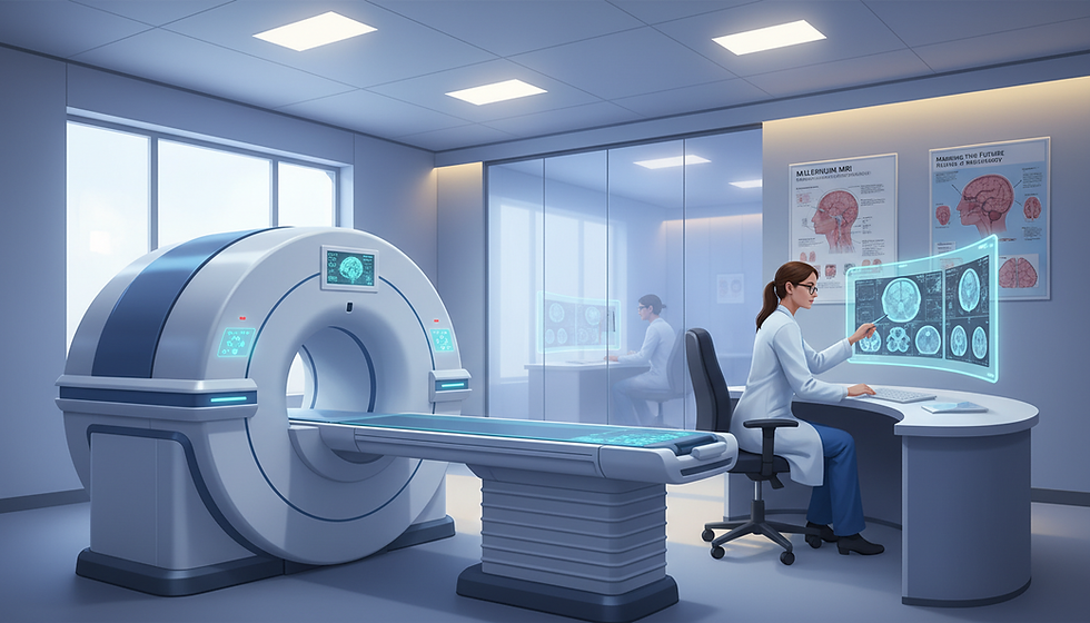
Magnetic resonance imaging (MRI) is a non-invasive medical imaging technique used to diagnose and treat a wide range of medical conditions. MRIs can detect abnormalities in soft tissues, organs, and bones, providing medical professionals with valuable information to guide treatment decisions. In this blog post, we will explore the capabilities of MRIs and the various conditions that they can detect.
I. Soft Tissue Injuries

MRIs are particularly effective at detecting soft tissue injuries, including muscle strains, ligament sprains, and tears. Soft tissue injuries can be challenging to diagnose using other imaging techniques, but MRIs can provide detailed images of the affected tissues, allowing medical professionals to identify the extent of the injury and develop an appropriate treatment plan.
Muscle strains: An MRI can detect tears, inflammation, and other injuries in the muscles. This information is important in developing an appropriate treatment plan and preventing further injury.
Ligament sprains: Ligament sprains can be difficult to diagnose with other imaging techniques, but an MRI can provide detailed images of the ligaments, allowing medical professionals to identify the extent of the injury and develop an appropriate treatment plan.
Torn tendons: Torn tendons can be detected on an MRI, allowing medical professionals to identify the extent of the injury and develop an appropriate treatment plan.
Rotator cuff injuries: An MRI can detect tears or other damage to the rotator cuff, allowing medical professionals to develop an appropriate treatment plan.
Meniscal tears: An MRI can provide detailed images of the knee joint and can detect meniscal tears, allowing medical professionals to develop an appropriate treatment plan.
Cartilage damage: An MRI can provide detailed images of the joint and can detect cartilage damage, allowing medical professionals to identify the extent of the injury and develop an appropriate treatment plan.
II. Brain Abnormalities

MRIs are frequently used to detect brain abnormalities, including tumors, aneurysms, and stroke. MRIs can provide detailed images of the brain and can detect abnormalities that may not be visible using other imaging techniques.
Brain tumors: MRI scans can detect tumors in the brain, including both cancerous and noncancerous tumors. MRI images can provide detailed information about the size, location, and type of tumor, which is important in developing an appropriate treatment plan.
Cerebral infarction: MRI scans can detect cerebral infarctions, which occur when blood flow to the brain is interrupted, leading to tissue damage. This type of damage can lead to cognitive deficits and motor impairment, so early detection is important.
Hemorrhage: MRI scans can detect hemorrhages in the brain, which occur when blood vessels rupture and cause bleeding. This type of bleeding can cause serious damage to the brain and can be life-threatening, so early detection is important.
Multiple Sclerosis: MRI scans are commonly used to diagnose multiple sclerosis (MS), a disease that affects the central nervous system. MRI images can detect the characteristic lesions that are indicative of MS, which is important in developing an appropriate treatment plan.
Aneurysms: MRI scans can detect aneurysms, which are bulges in the walls of blood vessels in the brain. Aneurysms can be life-threatening if they rupture, so early detection is important.
Stroke: MRI scans are commonly used to diagnose strokes, which occur when blood flow to the brain is interrupted. MRI images can provide detailed information about the location and extent of the stroke, which is important in developing an appropriate treatment plan.
Traumatic Brain Injury (TBI): MRI scans can detect traumatic brain injuries, which occur when the brain is damaged due to a blow to the head. MRI images can provide detailed information about the extent of the injury, which is important in developing an appropriate treatment plan.
III. Spinal Cord Injuries

MRIs can also detect spinal cord injuries, including herniated discs, spinal stenosis, and degenerative disc disease. MRIs can provide detailed images of the spinal cord and can identify abnormalities that may be causing pain or discomfort.
Herniated discs: MRI scans can detect herniated discs in the spine, which occur when the outer layer of the disc ruptures, causing the inner layer to protrude. This can cause pain, numbness, and weakness in the affected area.
Spinal cord tumors: MRI scans can detect tumors in the spinal cord, including both cancerous and noncancerous tumors. MRI images can provide detailed information about the size, location, and type of tumor, which is important in developing an appropriate treatment plan.
Spinal cord compression: MRI scans can detect spinal cord compression, which occurs when the spinal cord is compressed by a herniated disc, bone spur, or other abnormality. This can cause pain, numbness, and weakness in the affected area.
Spinal cord trauma: MRI scans can detect spinal cord trauma, which occurs when the spinal cord is damaged due to a blow to the back or neck. MRI images can provide detailed information about the extent of the injury, which is important in developing an appropriate treatment plan.
Spinal cord inflammation: MRI scans can detect inflammation in the spinal cord, which can be caused by a range of conditions, including infections and autoimmune disorders.
Spinal stenosis: MRI scans can detect spinal stenosis, which occurs when the spinal canal narrows, putting pressure on the spinal cord and nerves. This can cause pain, numbness, and weakness in the affected area.
IV. Bone Abnormalities
MRIs are also effective at detecting bone abnormalities, including fractures, tumors, and infections. MRIs can provide detailed images of the affected bone and can help medical professionals develop an appropriate treatment plan.
Fractures: MRI scans can detect fractures in the bones, particularly those that are not easily visible on X-rays.
Bone tumors: MRI scans can detect bone tumors, including both cancerous and noncancerous tumors. MRI images can provide detailed information about the size, location, and type of tumor, which is important in developing an appropriate treatment plan.
Osteoporosis: MRI scans can detect osteoporosis, a condition that causes bones to become weak and brittle. MRI images can provide information about bone density, which is important in assessing the severity of osteoporosis.
Arthritis: MRI scans can detect arthritis, a condition that causes inflammation in the joints. MRI images can provide information about the extent of joint damage, which is important in developing an appropriate treatment plan.
Bone infections: MRI scans can detect bone infections, which can be caused by bacteria or other microorganisms. MRI images can provide information about the extent of the infection, which is important in developing an appropriate treatment plan.
Stress fractures: MRI scans can detect stress fractures, which are small cracks in the bones that can be caused by repetitive stress or overuse.
V. Joint Injuries
MRIs are commonly used to detect joint injuries, including torn ligaments, meniscal tears, and cartilage damage. MRIs can provide detailed images of the affected joint, allowing medical professionals to identify the extent of the injury and develop an appropriate treatment plan.
Ligament injuries: MRI scans can detect tears, strains, and other injuries to ligaments, which are the tough bands of tissue that connect bones to one another. This information is important in developing an appropriate treatment plan and preventing further injury.
Tendon injuries: MRI scans can detect tears, inflammation, and other injuries to tendons, which are the fibrous tissues that connect muscles to bones. This information is important in developing an appropriate treatment plan and preventing further injury.
Meniscal tears: MRI scans can detect tears in the meniscus, which is the cartilage that acts as a cushion between the bones in the knee joint. This information is important in developing an appropriate treatment plan and preventing further injury.
Cartilage injuries: MRI scans can detect injuries to the cartilage in the joint, which can be caused by trauma or degenerative conditions such as arthritis. This information is important in developing an appropriate treatment plan and preventing further injury.
Joint effusions: MRI scans can detect joint effusions, which occur when excess fluid accumulates in the joint. This can be a sign of inflammation, infection, or other underlying conditions.
Synovitis: MRI scans can detect synovitis, which is inflammation of the synovial membrane that lines the joints. This can be a sign of arthritis or other inflammatory conditions.
VI. Abdominal and Pelvic Abnormalities
MRIs can also detect abnormalities in the abdominal and pelvic regions, including tumors, cysts, and infections. MRIs can provide detailed images of the affected organs, allowing medical professionals to identify the cause of the abnormality and develop an appropriate treatment plan.
Tumors: MRI scans can detect tumors in the abdominal and pelvic regions, including both cancerous and noncancerous tumors. MRI images can provide detailed information about the size, location, and type of tumor, which is important in developing an appropriate treatment plan.
Inflammatory bowel disease: MRI scans can detect inflammation in the digestive tract, which is a common sign of inflammatory bowel disease (IBD). MRI images can provide detailed information about the extent and severity of the inflammation, which is important in developing an appropriate treatment plan.
Gastrointestinal bleeding: MRI scans can detect sources of gastrointestinal bleeding, which can be caused by a range of conditions including ulcers, tumors, and inflammatory bowel disease.
Hernias: MRI scans can detect hernias, which occur when an internal organ or tissue pushes through a weak spot in the abdominal or pelvic muscles.
Endometriosis: MRI scans can detect endometriosis, a condition in which tissue that normally lines the inside of the uterus grows outside of the uterus. MRI images can provide detailed information about the location and extent of the endometrial tissue, which is important in developing an appropriate treatment plan.
Pelvic inflammatory disease: MRI scans can detect pelvic inflammatory disease (PID), which is an infection of the female reproductive organs. MRI images can provide detailed information about the extent and severity of the infection, which is important in developing an appropriate treatment plan.
VII. Cardiovascular Abnormalities
MRIs can detect abnormalities in the cardiovascular system, including heart disease, heart failure, and congenital heart defects. MRIs can provide detailed images of the heart and blood vessels, allowing medical professionals to identify the cause of the abnormality and develop an appropriate treatment plan.
Heart attacks: MRI scans can detect damage to the heart muscle caused by a heart attack. This information is important in developing an appropriate treatment plan and preventing further damage to the heart.
Coronary artery disease: MRI scans can detect blockages in the coronary arteries, which supply blood to the heart muscle. This information is important in assessing the severity of the blockages and developing an appropriate treatment plan.
Cardiomyopathy: MRI scans can detect cardiomyopathy, a condition in which the heart muscle becomes thickened or stiff. This information is important in developing an appropriate treatment plan and preventing further damage to the heart.
Congenital heart defects: MRI scans can detect congenital heart defects, which are abnormalities in the structure of the heart that are present at birth. This information is important in developing an appropriate treatment plan and preventing complications.
Aortic aneurysms: MRI scans can detect aortic aneurysms, which are bulges in the wall of the aorta. This information is important in assessing the size and location of the aneurysm and developing an appropriate treatment plan to prevent rupture.
Pericarditis: MRI scans can detect pericarditis, a condition in which the lining around the heart becomes inflamed. This information is important in developing an appropriate treatment plan and preventing complications.
VIII. Reproductive System Abnormalities
MRIs can also detect abnormalities in the reproductive system, including tumors, cysts, and fibroids. MRIs can provide detailed images of the affected organs, allowing medical professionals to identify the cause of the abnormality and develop an appropriate treatment plan.
Fibroids: MRI scans can detect fibroids, which are noncancerous growths in the uterus. MRI images can provide detailed information about the size, location, and type of fibroid, which is important in developing an appropriate treatment plan.
Ovarian cysts: MRI scans can detect ovarian cysts, which are fluid-filled sacs that form on the ovaries. MRI images can provide detailed information about the size, location, and type of cyst, which is important in developing an appropriate treatment plan.
Endometrial cancer: MRI scans can detect endometrial cancer, which is cancer that begins in the lining of the uterus. MRI images can provide detailed information about the extent and location of the cancer, which is important in developing an appropriate treatment plan.
Prostate cancer: MRI scans can detect prostate cancer, which is cancer that begins in the prostate gland. MRI images can provide detailed information about the extent and location of the cancer, which is important in developing an appropriate treatment plan.
Testicular cancer: MRI scans can detect testicular cancer, which is cancer that begins in the testicles. MRI images can provide detailed information about the extent and location of the cancer, which is important in developing an appropriate treatment plan.
Pelvic inflammatory disease: MRI scans can detect pelvic inflammatory disease (PID), which is an infection of the female reproductive organs. MRI images can provide detailed information about the extent and severity of the infection, which is important in developing an appropriate treatment plan.
IX. Dental and Maxillofacial Abnormalities
MRIs are frequently used to detect dental and maxillofacial abnormalities, including tumors, infections, and fractures. MRIs can provide detailed images of the affected area, allowing medical professionals to identify the cause of the abnormality and develop an appropriate treatment plan.
Temporomandibular joint (TMJ) disorders: MRI scans can detect problems with the TMJ, which is the joint that connects the jawbone to the skull. This information is important in developing an appropriate treatment plan and preventing further damage to the joint.
Dental infections: MRI scans can detect infections in the teeth and gums, which can be caused by bacteria or other microorganisms. This information is important in developing an appropriate treatment plan and preventing further infection.
Sinus infections: MRI scans can detect infections in the sinuses, which are air-filled spaces in the bones of the face. This information is important in developing an appropriate treatment plan and preventing further infection.
Tumors: MRI scans can detect tumors in the oral and maxillofacial region, including both cancerous and noncancerous tumors. MRI images can provide detailed information about the size, location, and type of tumor, which is important in developing an appropriate treatment plan.
Cleft lip and palate: MRI scans can detect cleft lip and palate, which are congenital abnormalities that occur when the tissues in the face and mouth do not fuse properly during fetal development. This information is important in developing an appropriate treatment plan.
Salivary gland disorders: MRI scans can detect disorders of the salivary glands, including tumors, infections, and blockages. This information is important in developing an appropriate treatment plan and preventing further damage to the glands.
X. Importance of Radiologist Expertise
Interpreting MRI images requires expertise and experience. Radiologists undergo extensive training and education to develop the skills needed to interpret medical images accurately. Their expertise is essential in ensuring that patients receive the correct diagnosis and treatment.
XI. Role of MRI in Cancer Detection and Treatment
MRIs play a significant role in detecting and treating cancer. They can detect tumors in various parts of the body, including the brain, breast, prostate, and liver. MRIs can provide detailed images of the tumor, allowing medical professionals to identify the size, location, and extent of the cancer. This information is crucial in developing an appropriate treatment plan.
In addition, MRIs are often used to monitor the effectiveness of cancer treatments, such as chemotherapy and radiation therapy. MRIs can provide detailed images of the tumor before and after treatment, allowing medical professionals to assess the effectiveness of the treatment and make any necessary adjustments.
XVII. Conclusion
In conclusion, MRIs are a powerful tool for medical diagnosis and treatment. They can detect a wide range of medical conditions, from soft tissue injuries to cancer, and provide detailed and accurate images of the body. The advancements in MRI technology continue to improve the accuracy and effectiveness of medical imaging, allowing for faster diagnoses and more targeted treatments. With greater accessibility and advances in technology, the future of MRI holds promise for even greater advancements in medical imaging.
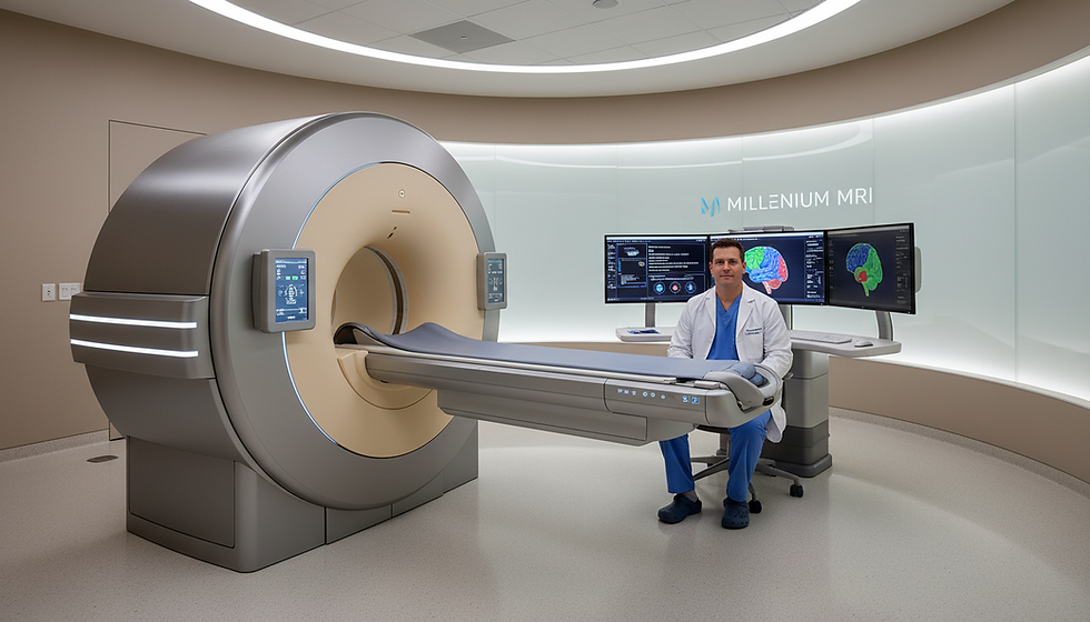
Schedule your MRI Today
Millennium MRI, with its state-of-the-art facilities situated in key locations including Largo, Florida; Tamarac, Florida; Ocala, Florida; Little Rock, Arkansas; Marion, Arkansas; and Jonesboro, Arkansas, has been at the forefront of offering advanced magnetic resonance imaging (MRI) diagnostic services. Their commitment to employing cutting-edge technology, coupled with a team of expert radiologists, ensures accurate and prompt results for patients. As one of the most trusted MRI centers in these regions, Millennium MRI prioritizes patient comfort and safety, making it the preferred choice for many seeking top-notch imaging services. Whether you're in the heart of Little Rock or the serene environs of Marion, know that Millennium MRI stands ready to serve your diagnostic needs.

Comments